
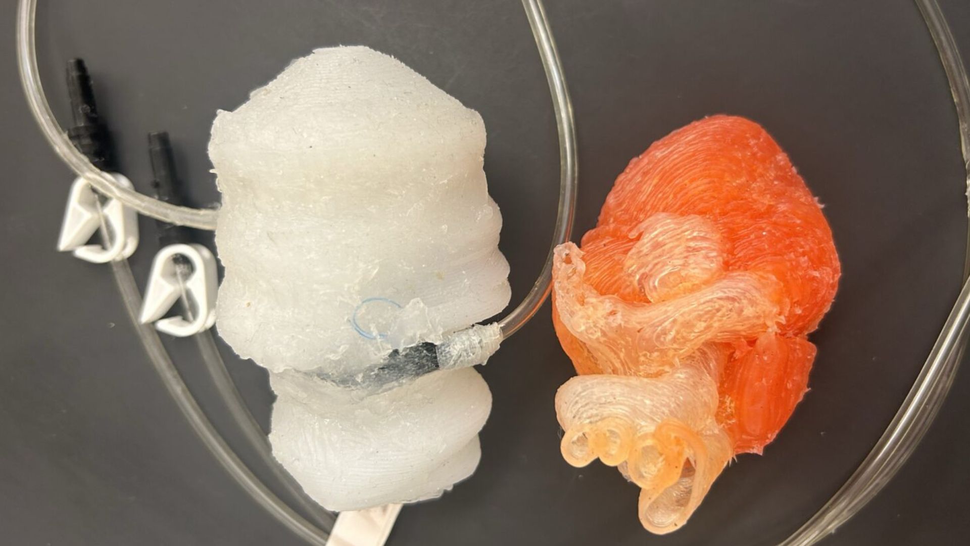
Researchers at Washington State University (WSU) have developed a 3D-printed heart model that can beat and contract.
The new model replicates the left side of the human heart, including the atrium, ventricle, and mitral valve. Scientists say it behaves much like a real heart and could improve surgical training and preparation for minimally invasive procedures.
The research findings were published in the journal Advanced Materials Technologies.
Doctors often need to perform delicate procedures on a beating heart. Practicing these techniques in a realistic setting is important for improving surgical accuracy.
Kaiyan Qiu, Berry Family Assistant Professor in WSU’s School of Mechanical and Materials Engineering and the lead researcher, said the new model offers a valuable training tool.
WATCH ALSO: https://modernmechanics24.com/wp-content/plugins/feeds-for-youtube/img/lightbox-placeholder.png
“It is very useful for doctors and surgeons to practice when the heart is still beating, especially for minimally invasive surgery,” Qiu said.
He added that the new model closely reproduces both the structure and movement of the heart.
“This model is the first fully synthetic system that mimics the complete left side of the heart without using animal models,” Qiu said.
Heart disease remains the leading cause of death in the US, and nearly 800,000 people undergo heart surgery every year.
Doctors commonly perform procedures to repair heart valves or treat other heart conditions. Traditionally, training for such procedures involves using animal models, cadavers, or computer simulations.
READ ALSO: https://modernmechanics24.com/post/iran-massive-drone-missile-attack-gulf/
However, these options have limitations. Animal and cadaver models cannot be reused and often do not represent the anatomy of individual patients.
Alejandro Guilllen Obando, a PhD candidate at WSU and the study’s first author, explained that many existing synthetic models also have design limitations.
“Most synthetic heart models are mold-casted, and they cannot reproduce the complex curves found in the human heart,” Obando said.
To create the model, the researchers used medical scans of a real human heart and converted them into a 3D-printed replica.
The team focused on the left side of the heart, which pumps oxygen-rich blood throughout the body. This area also experiences the highest blood pressure inside the heart.
READ ALSO: https://modernmechanics24.com/post/us-cyber-tools-sold-to-russia-for-years/
The model includes soft materials that mimic the texture of heart tissue. It also contains small pneumatic actuators that allow it to contract and beat like a real heart.
String-like components replicate the natural structures that control the movement of the mitral valve, which regulates blood flow between heart chambers.
Sensors attached to the model measure pressure as artificial blood flows through it. It allows researchers to monitor the heart’s behavior during simulated procedures.
To test the model, the research team created a damaged mitral valve and then performed a repair using a device similar to those used in hospitals.
WATCH ALSO: https://modernmechanics24.com/wp-content/plugins/feeds-for-youtube/img/lightbox-placeholder.png
During the procedure, the sensors recorded increased pressure inside the left ventricle, indicating that the valve was properly closing.
Ultrasound imaging also confirmed that the artificial blood was no longer leaking backward, demonstrating a successful repair.
The researchers have filed a provisional patent for their technology and plan to expand the project further. Their next goal is to create a complete heart model with all four chambers and four valves.
READ ALSO: https://modernmechanics24.com/post/china-space-mouse-gives-birth/
The team also hopes to work with doctors and medical students to use the model for patient-specific surgical rehearsals before real operations.
The project received funding from the National Science Foundation and from internal research and commercialization programs at WSU.
Researchers believe the technology could eventually help surgeons prepare more effectively and improve patient outcomes in heart surgery.


