
Scientists at UC Berkeley and Lawrence Berkeley National Laboratory have introduced a new cryo-electron microscopy technology that significantly improves images of small biological molecules.
The system combines an advanced laser phase plate with a specially designed microscope. The results were published in the journal Science.
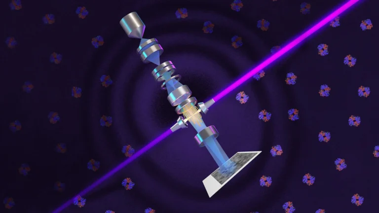
The new instrument is called Theia. Researchers designed it to capture sharper and clearer molecular images than conventional cryo-EM systems. The technology improves image contrast while preserving fine structural details.
Cryo-electron microscopy, commonly known as cryo-EM, is one of the most important tools in modern structural biology. It allows scientists to study proteins and other biological molecules at extremely small scales. The technique has played a major role in drug discovery and biomedical research.
Cryo-EM’s Small Molecule Problem
However, cryo-EM has limitations when studying smaller proteins. These molecules produce weak signals that are difficult to separate from background noise. As a result, scientists often need enormous amounts of data and extensive sample preparation.
The new laser phase plate directly addresses this challenge. It improves the visibility of molecular features that standard cryo-EM systems often struggle to detect. Researchers say the benefits become even more significant when imaging smaller proteins.
Project leader Holger Müller, a physics professor at UC Berkeley and faculty scientist at Berkeley Lab, described the impact in simple terms. He said traditional cryo-EM can feel like viewing artwork in a dark room. According to him, the new system effectively turns on the lights.
READ ALSO: General Atomics Sets Global Strategy for Swift Development of Uncrewed Combat Jets
How the Phase-Contrast Technology Works
The technology is inspired by a concept first developed for light microscopy nearly a century ago. In the 1930s, Dutch physicist Frits Zernike discovered a method to improve contrast in transparent biological samples. His invention later earned the 1953 Nobel Prize in Physics.
Cells are often difficult to see under normal microscopes because they are largely transparent. Light passing through them changes slightly in phase rather than brightness. Zernike found a way to convert those invisible phase changes into visible image contrast.
Scientists attempted to apply the same principle to electron microscopy for decades. Many experimental phase plates either weakened the electron beam, reduced image quality, or introduced instability. These limitations prevented widespread adoption.
In 2010, Müller and cryo-EM pioneer Robert Glaeser began working on a new solution. Instead of using traditional phase plates, they proposed using an extremely powerful laser. Their goal was to shift the phase of electron waves without damaging image quality.
Creating the required laser system took more than a decade of research and engineering. The team built a mirrored optical cavity that traps and intensifies laser light. As the light reflects thousands of times, its power increases dramatically.
The final system generates an extraordinary level of brightness. Researchers say the laser delivers about 75 kilowatts of power concentrated into an area only a few microns wide. According to the team, this produces the brightest continuous laser focus ever created.
Theia Shows Strong Results on Difficult Samples
To test the technology, researchers imaged two well-known proteins. One was aldolase, a protein found in muscle tissue. The second was hemoglobin, the oxygen-carrying protein found in blood.
READ ALSO: Westminster Abbey discovers link with Charlemagne
Aldolase is relatively easy to study using existing cryo-EM systems. Hemoglobin is much smaller and represents a more demanding test. It sits near the lower size limit of what current cryo-EM technology can reliably analyze.
The laser phase plate improved image quality for both proteins. However, the gains were especially noticeable for hemoglobin. The results suggest the system performs best when imaging molecules that are traditionally difficult to capture.
Researchers also tested samples with varying levels of quality. Some samples were carefully prepared, while others contained imperfections that normally reduce imaging performance. The new system consistently improved results across different conditions.
Sample preparation remains one of the most time-consuming parts of cryo-EM research. Scientists must freeze thousands, or even millions, of molecules at temperatures below -160 degrees Celsius. Even tiny amounts of ice contamination can interfere with imaging.
The new technology helps reduce some of these challenges. Producing a stronger contrast allows researchers to extract more useful information from difficult samples. This can potentially save time and improve research efficiency.
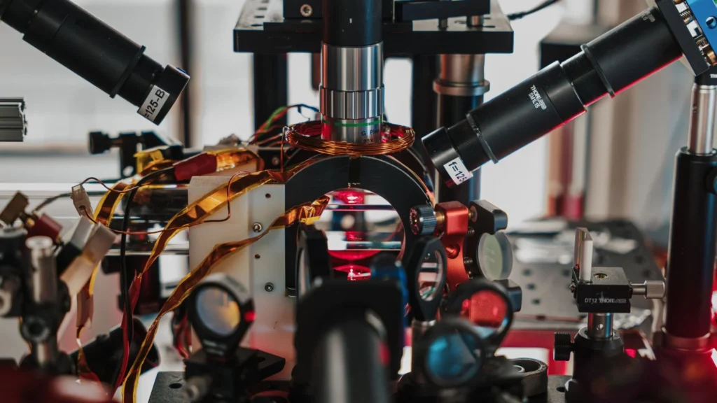
The complete Theia system was finalized and installed in 2025. It combines the laser phase plate with a customized Thermo Fisher Scientific microscope. Within days of installation, the team began capturing highly detailed images.
The project received support from multiple organizations. Funding came from the National Institutes of Health, the National Science Foundation, the Gordon and Betty Moore Foundation, the Chan Zuckerberg Initiative, and Berkeley Lab’s internal research programs.
READ ALSO: China’s 3D-Printed Mini Jet Engine Succeeds in Historic Flight
Expanding Toward 3D Imaging Inside Cells
Researchers are now preparing to extend the technology to cryo-electron tomography (cryo-ET). This technique creates three-dimensional views of molecules and cellular structures. It works by combining images captured from multiple angles.
Cryo-ET offers an important advantage over traditional single-particle analysis. It allows scientists to observe molecules inside cells rather than studying isolated samples. This provides a more realistic picture of how biological systems function.
Interpreting cryo-ET data can be difficult because cellular environments are crowded and complex. Low signal levels often make important structures hard to identify. The laser phase plate is expected to improve visibility in these challenging conditions.
Jessie Zhang, a postdoctoral researcher involved in the study, said the technology can help scientists observe more proteins in their natural environment. Better image quality could reveal biological processes that remain hidden today. This would expand understanding of how cells operate in real time.
The research team is also working to improve the microscope’s focusing capabilities. Current cryo-EM systems intentionally use slightly defocused electron beams to generate contrast. Because the laser phase plate already provides strong contrast, future versions may operate with sharper focus.
Scientists estimate this adjustment could double the amount of useful structural information captured in each image. Such improvements would push cryo-EM closer to imaging even smaller proteins. The long-term target is to study molecules weighing around 17 kilodaltons.
That goal is particularly important because most human proteins are relatively small. Researchers estimate that about 90 percent of human proteins fall below the size range that current cryo-EM systems handle comfortably. Expanding imaging capabilities would therefore benefit a vast area of biological research.
WATCH ALSO: Apple launches iPhone 17 lineup with contoured edges, and thinner borders
Theia remains a prototype system today, but researchers are already exploring ways to make the technology more accessible. Collaborations with microscope manufacturers aim to develop versions that are easier to operate. These future systems could eventually be installed in research centers worldwide.
The successful integration of laser phase-contrast imaging into cryo-EM represents a major step in biological imaging technology. By delivering clearer views of tiny proteins and improving the study of molecules inside living systems, the new platform gives scientists a stronger tool for exploring the machinery of life.