Scientists at Novosibirsk State University have discovered evidence of advanced jaw surgery in a woman who lived more than 2,500 years ago.
The woman belonged to the ancient Pazyryk culture, a nomadic society known for its well-preserved frozen tombs in the Altai Mountains. Researchers say the findings reveal a level of medical skill not previously linked to Iron Age communities in Siberia.
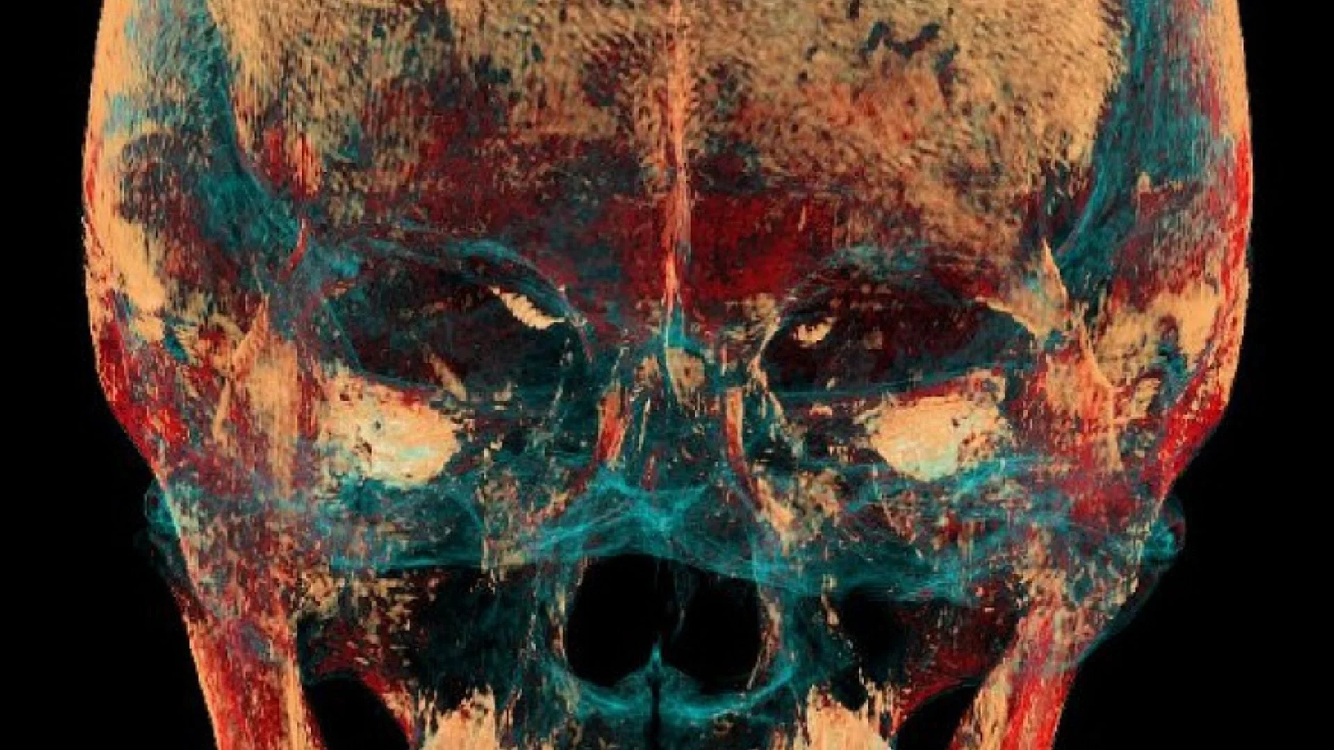
The discovery came from a detailed CT scan of the woman’s skull. Scientists found signs of surgical drilling and jaw stabilization after a severe injury to the right side of her face. The operation appears to have restored her ability to eat and speak, allowing her to survive for a long period after the trauma.
The woman was buried at the Verkh-Kaldzhin-2 burial ground on the Ukok Plateau in Russia’s Altai Republic. Archaeologists first discovered the site in 1994 during excavations connected to the Pazyryk culture. The frozen burial conditions helped preserve human remains, textiles, leather, and other organic materials for centuries.
Researchers estimated the woman was between 25 and 30 years old at the time of her death. Her skull remained partly covered with preserved soft tissue, which made earlier examinations difficult. Modern imaging technology finally allowed experts to examine the hidden structures without damaging the remains.
READ ALSO: CIA’s Lost Plutonium Device on Nanda Devi Still Haunts Uttarakhand After 60 Years
Scientists from the Laboratory of Nuclear and Innovative Medicine at NSU carried out the scan using a Philips MX 16 CT scanner. The system produced 551 thin-slice images of the skull, creating a detailed three-dimensional digital model. Researchers described the technology as a ‘time machine’ because it enabled them to study injuries and treatments that had been hidden for centuries.
Ancient Jaw Surgery Revealed
The scans revealed a serious injury to the right temporal bone near the jaw joint. A deep depression in the bone had damaged the temporomandibular joint(TMJ). The injury displaced the lower jaw and tore supporting ligaments that normally help the jaw move properly.
Doctors today consider this type of injury extremely dangerous without treatment. Severe jaw damage can prevent a person from chewing, swallowing, or speaking normally. In ancient times, such trauma often led to infection, starvation, or death.
The woman’s teeth showed clear evidence that she survived long after the injury occurred. Researchers found heavy wear on the left side of her mouth, while the right side remained less damaged. This suggested she relied mainly on the uninjured side of her jaw for chewing over an extended period.
CT images also uncovered two carefully drilled bone canals near the damaged joint. The canals measured about 1.5 millimeters wide and intersected at a right angle. Researchers said the smooth drilling marks proved the holes were created intentionally rather than caused by the injury itself.
The bone around the drilled areas showed signs of healing. This confirmed the procedure was performed while the woman was alive and that she survived afterward. Scientists also identified traces of soft material inside the canals, possibly horsehair or animal tendon.
Researchers believe ancient surgeons used the material as a surgical ligature. The fibers appear to have helped stabilize the jaw and hold damaged joint structures in place. In simple terms, the procedure worked like an early fixation system similar to techniques used in modern reconstructive surgery.
Radiologist Andrey Letyagin said the operation restored jaw joint movement. Although chewing on the injured side remained painful, the surgery helped the woman regain important daily functions. Researchers believe this treatment gave her a chance to continue living despite the severe trauma.
Discovery Changes Understanding of Iron Age Medicine
The findings are important because scientists have not previously documented this type of surgery in the Pazyryk culture. Earlier research had already shown that the society practiced skull trepanation and complex mummification methods. The new evidence adds orthopedic-style surgery to the list of their medical abilities.
The Pazyryk people lived between the 6th and 3rd centuries BC across parts of Siberia and Central Asia. They belonged to the wider Scythian world and were skilled horse riders, traders, and metalworkers. Their frozen tombs have become famous for preserving tattoos, clothing, wooden objects, and even mummified bodies.
Researchers believe mummification practices helped ancient healers learn more about human anatomy. Preparing bodies through dissection gave them direct knowledge of muscles, bones, and organs. That understanding may have supported the development of surgical techniques within the community.
Scientists still do not know exactly how the woman suffered the injury. Experts say it may have resulted from a horse-riding accident, a fall, or a violent blow to the head. The Altai region’s harsh environment and physically demanding lifestyle increased the risk of serious injuries.
Life in the ancient mountain valleys was difficult and dangerous. Small populations depended heavily on every member of the community for survival. Researchers say this may explain why people invested time and skill into difficult medical procedures instead of abandoning badly injured individuals.
The discovery also highlights the growing role of modern imaging technology in archaeology. CT scanners allow scientists to examine fragile remains without cutting or damaging them. High-resolution scans can reveal hidden fractures, internal healing, and surgical marks that traditional methods sometimes miss.
Unlike medical scans used for living patients, archaeologists can use higher X-ray settings to capture more detailed images. This helps researchers study ancient bones with greater precision. In this case, the extra detail exposed the tiny drilled canals and traces of healing tissue.
WATCH ALSO: Chinese company’s humanoid robot has mastered Webster flip, defies physics
The findings challenge older ideas about the limits of ancient medicine. Many people assume advanced surgery developed only in later civilizations with formal medical systems. The Pazyryk evidence shows that practical surgical knowledge also existed in remote Iron Age societies.
The study also shows that ancient communities valued care and recovery. Performing such a difficult operation required planning, tools, and specialized knowledge. Researchers say the woman’s survival reflects both medical skill and social support within her community.
Experts believe similar discoveries may still be hidden inside ancient remains stored in museums and research collections. New scanning technology is helping scientists reexamine old finds with fresh detail. Each new discovery adds more evidence that ancient societies possessed deeper medical knowledge than once believed.
The Pazyryk woman’s skull now stands as one of the clearest examples of ancient surgical treatment in Siberia. Through modern CT imaging, researchers uncovered a story of injury, treatment, and survival that remained hidden for 25 centuries. Scientists say future research into ancient medicine may continue to reshape what the world knows about early human healthcare and surgical innovation.