

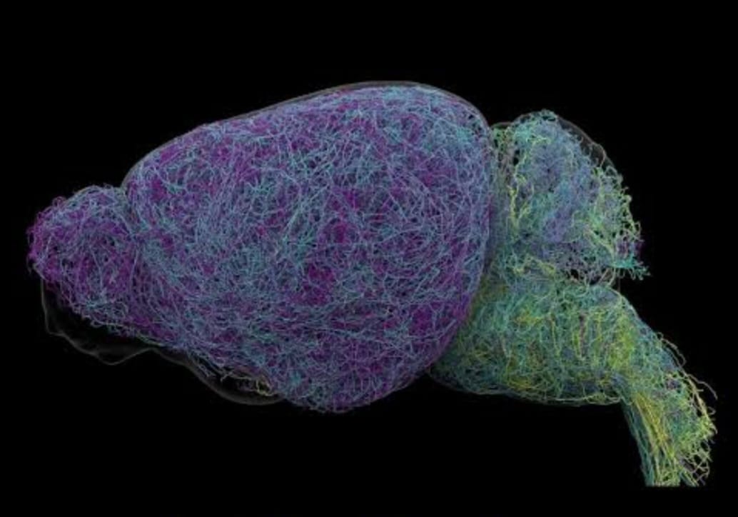
Scientists have produced a detailed map of the mouse brain’s locus coeruleus, a small region that controls the release of the chemical messenger norepinephrine.
The study shows that its neurons do not send one broad signal across the brain, as previously thought. Instead, different groups of neurons connect to specific brain regions and appear to support different functions, including learning and attention.
The locus coeruleus (LC), sits deep inside the brainstem and contains only a small share of the brain’s neurons. Its name means “blue place” in Latin, referring to the region’s appearance under certain staining methods. The LC releases norepinephrine (NE) which helps regulate attention, learning, stress responses, heart rate and other body functions.
For years, scientists often described the LC as a system that broadly releases NE across the brain. The new research presents a more detailed picture of how these signals travel. Allen Institute researchers combined brain imaging, neural recordings, genetic analysis and behavioral experiments to study the connections and functions of LC neurons in mice.
READ ALSO: https://modernmechanics24.com/post/tkms-atlas-uk-royal-navy-torpedo-contract/
The researchers found a clear difference between the upper and lower parts of the LC. Neurons in the dorsal, or upper, LC mainly sent their long connections toward the cerebral cortex and forebrain. Neurons in the ventral, or lower, LC mainly connected with the brainstem and spinal cord.
Karel Svoboda, director of Neural Dynamics at the Allen Institute and a co-author of the study, compared the system with a targeted postal network rather than a foghorn. The comparison describes how different LC neurons send signals to particular destinations instead of releasing the same message throughout the brain.
Neurons Linked To Learning
The researchers also examined how LC neurons responded while mice performed decision-making tasks. Some of the tasks required the animals to change their choices after receiving a negative result. During these situations, dorsal LC neurons that connect toward the cortex became more active.
The team found a different pattern in ventral LC neurons. These cells showed increased activity shortly before mice ignored cues that might have indicated a possible reward. The findings link different LC pathways with separate aspects of behavior.
Svoboda said the researchers identified a pattern in which dorsal LC neurons that project toward the cortex are involved in learning. Ventral neurons that project downward toward the brainstem and spinal cord were associated with whether the animals engaged with their surroundings.
The researchers also found differences in gene activity between these groups of neurons. This means that the anatomical differences were accompanied by distinct genetic profiles. The results provide a more detailed view of how the LC is organized and how its different cell populations operate.
WATCH ALSO: https://modernmechanics24.com/post/nasa-roman-telescope-new-view-universe/
Longest Mouse Neuron Found
The study involved extensive mapping of the mouse brain. Researchers used whole-brain imaging to examine nearly 35,000 neurons and genetic profiling to study almost 400,000 cells.
They also reconstructed some of the brain’s most complex neurons in detail. One of the most notable findings involved the length of their axons. An axon is a long extension of a neuron that carries electrical signals toward other cells.
The LC neurons examined in the study had axons averaging about 35 centimeters in length. One measured 70.32 centimeters, making it the longest single neuron measured in a mouse, according to the researchers.
Jeremiah Cohen, an Allen Institute scientist and co-author, said the neuron supplied norepinephrine to a large area of the cerebral cortex. Despite its wide reach, the neuron did not release NE throughout the entire brain.
Cohen said the neuron bypassed the cerebellum, brainstem and spinal cord. This finding shows that even neurons with very long projections can have defined targets.
Implications For Brain Medicine
Norepinephrine is involved in attention, mood and learning. Because of these functions, the chemical system is affected by medicines used to treat conditions such as depression, anxiety and attention-deficit/hyperactivity disorder.
The LC also has a connection with neurodegenerative disease research. LC neurons are among the earliest groups of neurons affected by degeneration in Alzheimer’s disease. A clearer map of their connections may therefore help researchers understand how changes in these cells affect brain function.
The study, published in Nature, also examined how norepinephrine interacts with dopamine. Dopamine is another major chemical messenger in the brain and sends signals to areas such as the basal ganglia, which is involved in habit formation.
The researchers propose that norepinephrine and dopamine systems work together as part of a learning network. Their findings suggest that these systems may help the brain process different forms of learning and behavior at the same time.
The detailed map may also help researchers study whether future treatments can act on particular neural pathways. Instead of affecting the entire norepinephrine system, therapies may eventually be designed around specific circuits, although such applications require further research.
READ ALSO: https://modernmechanics24.com/post/germany-lockheed-martin-f35a-fighter/
The research was supported by the National Institutes of Health through its Brain Research Through Advancing Innovative Neurotechnologies Initiative, known as the BRAIN Initiative. By combining large-scale imaging, genetic profiling and behavioral studies, the work provides a detailed picture of how a small brain region sends different signals to different parts of the nervous system.
The findings also give researchers a clearer framework for studying how norepinephrine supports learning and behavior. Further work will be needed to determine how closely these pathways in mice correspond to the human brain and whether the new anatomical information can lead to more precise treatments for neurological and psychiatric conditions.














One Response