

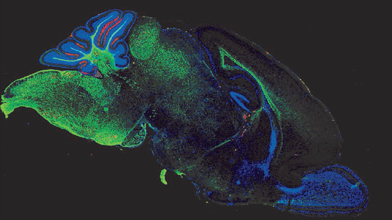
Researchers at Stanford University have developed mice containing human brain tissue that occupies about half the volume of their brains.
The study gives scientists a new way to examine neurological and psychiatric disorders inside living animals, including conditions such as schizophrenia, epilepsy, cerebral palsy, intellectual disability and rare forms of dementia.
The research, published in Nature, also raises questions about animal welfare and the limits of human brain organoid experiments.
The team transplanted laboratory-grown human brain cells into genetically engineered mice that were born without two major brain regions. These regions, the cerebral cortex and hippocampus, normally support functions such as movement, memory, learning and processing information. Their absence created space for human brain tissue to develop inside the animals’ skulls.
The approach addresses a long-standing challenge in brain research. Scientists can study human cells in laboratory dishes, but these models do not fully reproduce the conditions of a living brain. By placing human tissue inside animals, researchers can examine how it develops, connects with other neurons and responds to disease-related changes.
READ ALSO: https://modernmechanics24.com/post/study-find-hidden-reactor-tube-stresses/
Creating Space for Human Tissue
The Stanford research was led by Sergiu Pașca, a professor of psychiatry. His team has been investigating ways to make human brain development and function more accessible for scientific study.
The researchers began with mice that had been genetically modified to lack the cerebral cortex and hippocampus. These regions normally occupy a substantial part of the brain, but the modified animals survived because other parts of their nervous systems adapted to perform additional functions.
The mice appeared broadly normal. However, they showed a cautious gait and memory-related difficulties, reflecting the effects of losing important brain structures.
The scientists then introduced human brain organoids into the developing mice. Organoids are small, three-dimensional clusters of cells grown in laboratories from stem cells or reprogrammed adult cells. They can reproduce some features of human brain tissue, although they do not replicate the full structure or function of a human brain.
The human cells came from donated skin cells. Researchers reprogrammed those cells into a stem-cell-like state and used them to produce brain tissue in the laboratory.
The newborn mice received several injections, each containing approximately 100,000 human brain cells. The cells were placed in the space where the animals’ own brain tissue was missing.
The mice lacked around 14 million mouse brain cells in total. After transplantation, they developed approximately four million human brain cells, with the human tissue accounting for about half of the brain’s volume.
How Human Neurons Connect
Three months after transplantation, the human tissue had almost completely filled the cavity created by the missing brain regions. It had also established a blood supply from the mouse, allowing the transplanted tissue to receive oxygen and nutrients.
Some human neurons formed connections with mouse brain cells. Other connections extended toward parts of the spinal cord, showing that the transplanted tissue was able to interact with the animals’ existing nervous systems.
The human tissue did not develop into a normal human brain. Its organization and wiring differed from those found in people, and the neurons remained immature. The researchers estimated that the tissue resembled the developmental stage of a human brain around the middle of pregnancy.
The scientists called the animals ‘xenocortical’ mice. The name refers to the presence of human cortical tissue inside a different species.
WATCH ALSO: https://modernmechanics24.com/post/china-c919-first-international-flight/
Tests showed that the transplants did not enhance the animals’ abilities. The mice did not become more intelligent or acquire human-like cognitive skills. However, their shaky gait and some of their cognitive difficulties improved slightly after the human tissue developed.
The findings suggest that human neurons can function within an animal brain without producing a human mind. They also show that transplanted tissue may influence the activity of surrounding neural circuits.
Studying Disease in Living Brains
One of the study’s main goals is to create a model for investigating human neurological disorders. Researchers can take cells from patients, grow brain tissue from those cells and place it inside living animals. This offers a way to examine disease-related changes in human neurons under biological conditions that laboratory dishes cannot fully reproduce.
The team used oxygen deprivation to demonstrate the model’s potential. Some mice were exposed to five hours of low oxygen, a condition that can damage developing human brain tissue.
Oxygen deprivation during pregnancy or childbirth is associated with certain cases of cerebral palsy. The researchers found that the human brain tissue in the mice was vulnerable to this stress, providing an opportunity to study how oxygen loss affects human nerve cells.
The model may also help scientists investigate conditions such as schizophrenia, epilepsy and intellectual disability. These disorders involve complex changes in brain development or function, and studying patient-derived human tissue may reveal mechanisms that are difficult to observe directly in people.
The researchers also identified rare cells called von Economo neurons in the transplanted tissue. These specialized neurons have been observed in humans and certain other large-brained mammals, but they are difficult to study because researchers generally identify them through postmortem brain examinations.
Von Economo neurons are among the cells that are lost early in frontotemporal dementia, a rare form of dementia that affects behavior, personality and language. Pașca hopes to use the xenocortical mice to investigate how these neurons develop and why they may be vulnerable to disease.
READ ALSO: https://modernmechanics24.com/post/byd-62mwh-gc-block-grid-storage/
Ethics and Future Research
The research belongs to the expanding field of human neural organoids. Scientists use these models to study brain development, disease mechanisms and potential treatments without relying exclusively on human subjects or animal experiments.
However, organoid research also presents ethical questions. Researchers and bioethicists are examining whether increasingly complex brain tissue might develop forms of awareness or the capacity to experience pain. The welfare of animals that receive human brain tissue is another important concern.
Pașca said the Stanford work received extensive ethical oversight from the beginning. He argued that the research aims to address the limited availability of effective treatments for many psychiatric and neurological conditions.
Emily Jackson, a professor of law at the London School of Economics and chair of a recent Nuffield Council on Bioethics report on neural organoids, emphasized the need for continued monitoring. She said researchers must closely observe the animals to evaluate the effects of the transplanted tissue on their welfare.
Madeline Lancaster, a group leader at the MRC Laboratory of Molecular Biology in Cambridge, said the approach may be useful for studying disorders and treatments that require a whole animal.
She questioned whether it would provide a complete picture of normal human brain development because the model is artificial and does not follow the natural process of human brain formation.
Lancaster also noted that many researchers are developing brain organoids entirely in laboratory dishes. These systems may help reduce the number of animals used in research, although they still face challenges in reproducing the maturity and complexity of human brain tissue.
The Stanford study therefore represents one part of a broader effort to improve disease models. Its value will depend on how accurately the transplanted tissue reproduces human disease and whether the findings lead to treatments that work in patients.
For now, the xenocortical mice provide researchers with a new way to study human neurons in a living biological environment. Further work will determine how far this model can advance understanding of brain disorders while maintaining appropriate ethical safeguards.














One Response