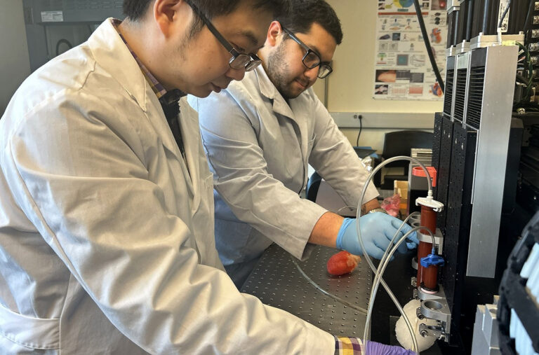
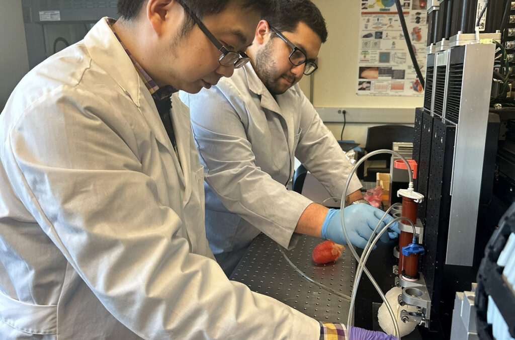
Researchers at Washington State University have created a soft, synthetic left heart model that pumps and beats like the real organ. Surgeons can practice valve repairs on it using real tools and imaging.
A team at Washington State University has developed a 3D-printed model of the left side of the heart that actually contracts and beats. The researchers successfully performed a mitral valve repair on the model, using ultrasound imaging and sensors to confirm the fix worked. The work was published in the journal Advanced Materials Technologies.
Kaiyan Qiu, Berry Family Assistant Professor in the School of Mechanical and Materials Engineering, led the research as corresponding author. Alejandro Guillen Obando, a PhD candidate in the same school, is first author. The team used a scan of a real human heart to build their replica, focusing on the left side where pressure is highest and vital pumping happens.
READ ALSO: https://modernmechanics24.com/post/durapatcher-technology-pothole-crisis/
Heart disease is the leading cause of death in the United States, with about 800,000 heart surgeries performed each year. Training for these procedures often means practicing on animals or cadavers, which are not reusable and do not match individual patients perfectly. Computer simulations offer another option, but they lack hands-on realism. A synthetic model that behaves like living tissue could fill this gap.
The model includes the left atrium, left ventricle, and the mitral valve between them. It is made from soft materials that feel similar to real heart tissue. Tiny pneumatic actuators make the model pump, while string-like structures control valve movement. Sensors track imitation blood pressure as fluid flows through. The layer-by-layer 3D printing method allows complex curves that molded models cannot achieve.
To test their creation, the team printed a defective mitral valve and repaired it using a device similar to commercial tools. Sensors showed blood pressure in the left ventricle increased after repair, meaning the valve closed properly. Ultrasound images confirmed that imitation blood stopped flowing backward. This proves the model can simulate both healthy and diseased hearts for practice.
The current model only covers the left side of the heart. The team is now working on a full version with all four chambers and four valves. They also plan to collaborate with medical professionals to conduct patient-specific rehearsals for different valve diseases. A provisional patent has been filed.
WATCH ALSO: https://modernmechanics24.com/post/china-40000t-sichuan-assault-ship/
This model offers a reusable, realistic training tool that could reduce dependence on animals and cadavers. Surgeons could practice difficult procedures beforehand on a replica of a specific patient’s heart. The work was funded by the National Science Foundation and WSU internal grants, showing strong interest in improving surgical outcomes through better training tools.