Doctors in China have used a microwave ablation needle to shrink a large lung tumour in an unborn baby, treating the growth while the foetus was still inside the womb.
The procedure was performed at 22 weeks of pregnancy and helped prevent further complications caused by the rapidly growing tumour. The baby was later delivered at 37 weeks and remained healthy at four months.
The case involved a 40-year-old woman whose foetus was diagnosed with microcystic congenital pulmonary airway malformation (CPAM) at 22 weeks of pregnancy.
CPAM is a rare condition in which abnormal lung tissue develops during pregnancy. Although the condition is benign, some forms can grow rapidly and place severe pressure on the developing heart and lungs.
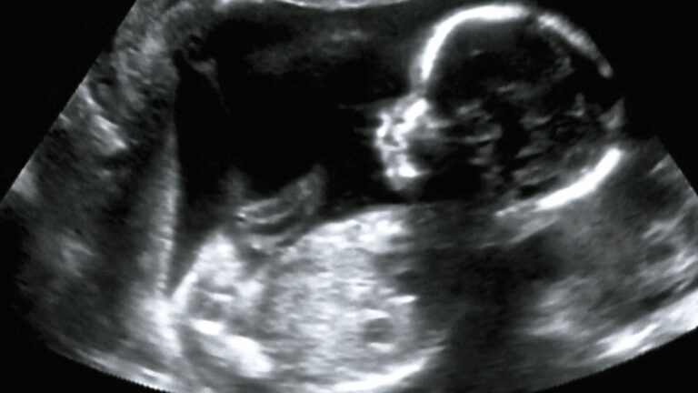
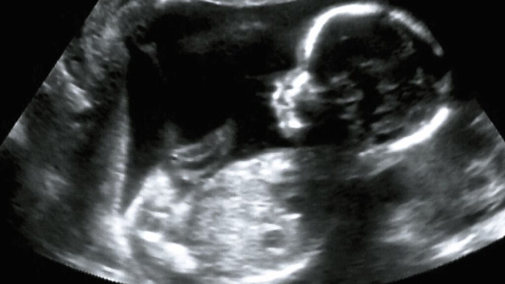
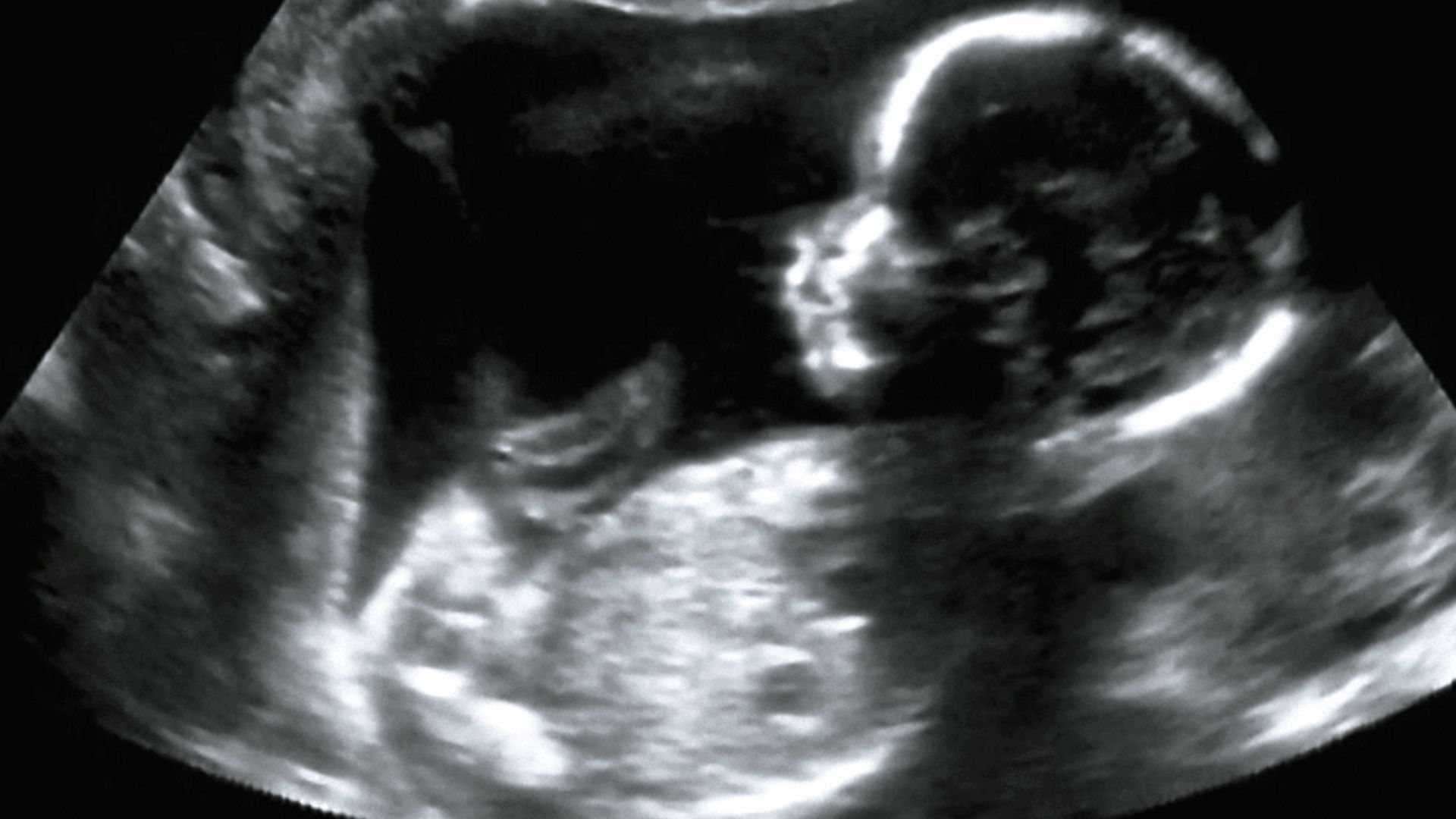
Ultrasound scans showed a solid mass measuring about 5cm in the foetus’ right chest cavity. The tumour filled much of the space and pushed against the heart and lungs. The pressure had already caused ascites, a condition in which fluid builds up inside the abdomen.
Large CPAMs can become dangerous before birth because they take up space needed by the developing lungs and heart. In severe cases, the condition can lead to hydrops, which involves abnormal fluid accumulation in several parts of the foetus. Early treatment is therefore important when the tumour continues to grow and affects vital organs.
Why Standard Treatment Failed
Doctors normally have several options for treating severe foetal lung lesions before birth. These include draining fluid-filled cysts or blocking the blood vessel supplying the tumor. Those approaches were unsuitable in this case because the lesion consisted mainly of very small cysts and lacked a single major feeding blood vessel.
The medical team therefore chose a different method. Doctors from Shenzhen Baoan Women’s and Children’s Hospital worked with specialists from the Chinese University of Hong Kong on the procedure. The case was reported in the peer-reviewed journal Ultrasound in Obstetrics & Gynecology on August 20.
The team used high-definition ultrasound to plan the procedure in real time. This allowed doctors to identify the tumor and choose a path that reduced risk to the fetus and surrounding maternal tissue. The approach also allowed doctors to monitor the treatment as it took place.
READ ALSO: https://modernmechanics24.com/post/us-australia-hacm-hypersonic-missile/
Microwave Energy Targets Tumour
A thin ablation needle was inserted through the mother’s abdomen and uterine wall before entering the foetal chest. Doctors guided the needle directly into the centre of the tumour using ultrasound imaging. The procedure was performed without opening the uterus through conventional surgery.
The needle delivered microwave energy into the tumour. The energy causes water molecules in the tissue to move rapidly, producing heat that damages and destroys the targeted cells. In this case, the active heating stage lasted about two minutes.
Doctors closely monitored the treatment to control where the heat traveled. The aim was to destroy the abnormal lung tissue while protecting the foetus’ developing organs. The immediate response suggested that the treatment had reduced the pressure caused by the tumour.
Baby Reaches Full-Term Delivery
Three days after the procedure, scans showed a major reduction in the tumour’s size. The fluid accumulation in the foetus’ abdomen had also disappeared. The pregnancy was then allowed to continue under medical monitoring.
The baby was delivered by caesarean section at 37 weeks. The newborn weighed 2.9kg and received an Apgar score of 10, indicating a strong condition immediately after birth. The tumor did not require immediate surgical treatment.
At four months of age, the infant remained symptom-free. The baby had not needed further surgery during that period. The reported outcome provides a clinical example of how image-guided treatment can be used for selected foetal tumours before birth.
The Shenzhen and Hong Kong medical teams jointly reported the procedure, and Chinese science media described it as a new milestone in minimally invasive treatment for solid fetal tumors. The case also adds to research on treating serious foetal conditions without conventional open foetal surgery.
For maternal-foetal medicine, the case points to a wider role for precisely targeted treatments during pregnancy. More clinical experience is needed to determine how broadly microwave ablation can be used for similar foetal tumors and which patients are most suitable for the approach.