MIT engineers have developed a paper-thin swimming robot powered by a single layer of living muscle cells.
The biohybrid machine uses light-responsive muscle tissue attached to a thin gel structure to generate movement in water.
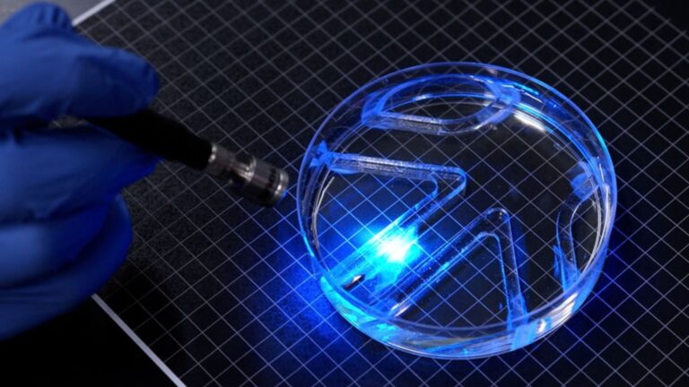
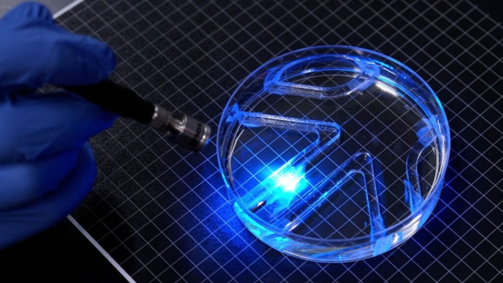
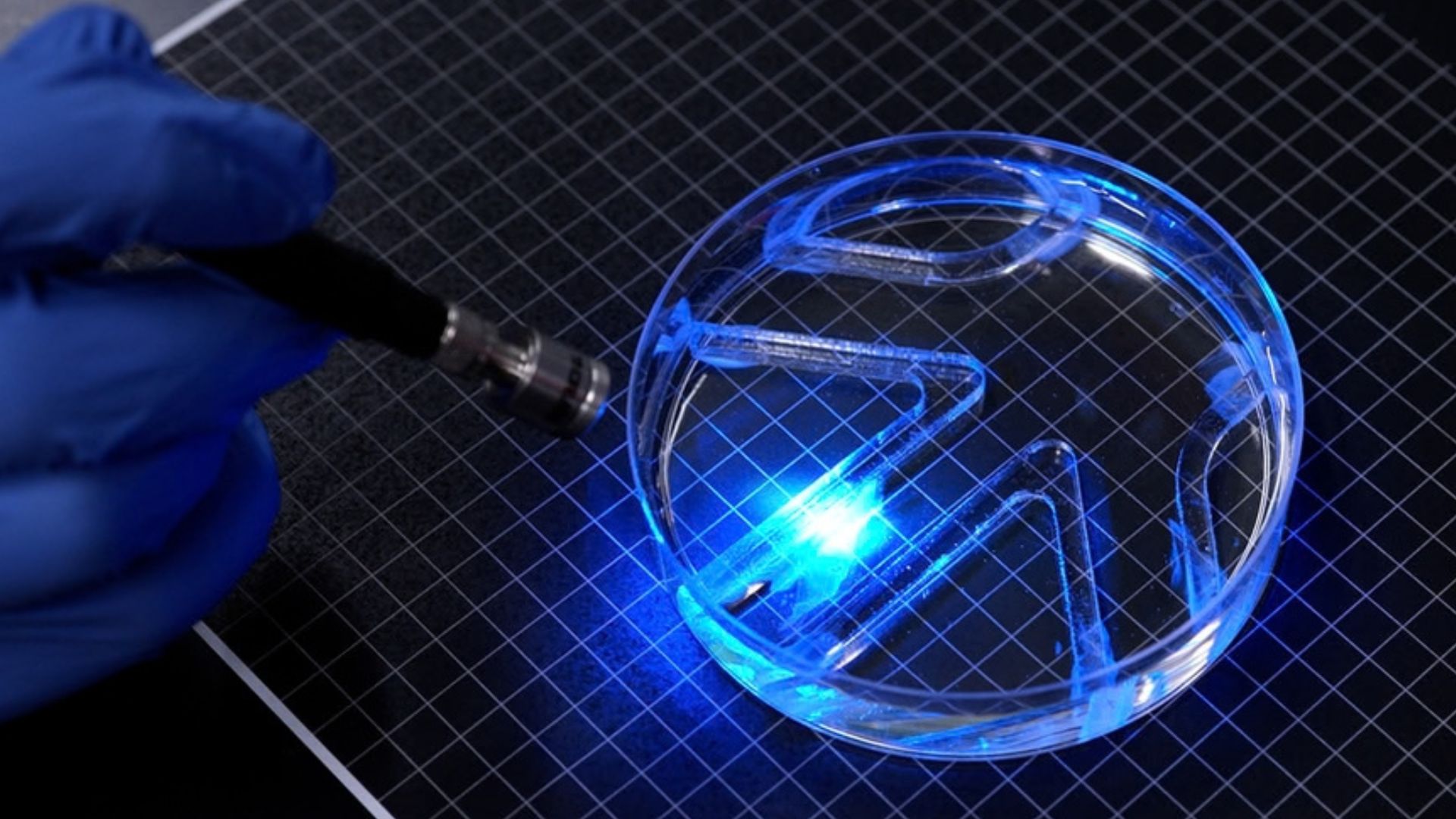
Researchers demonstrated the robot swimming and turning through a small aquatic maze, showing how living tissue can drive compact robotic systems.
The study appears in Advanced Functional Materials. It presents a two-dimensional design that is much thinner than many earlier muscle-powered robots.
The work also explores how the material supporting the muscle cells affects their alignment, strength and ability to move a robot.
The new aquabot is roughly the length and width of a stick of gum. Its main structure is a thin film of gel divided into two sections that act as fins. Each fin carries a layer of living skeletal muscle cells that is thinner than a strand of human hair.
The cells were genetically engineered to respond to light. When researchers shine light on one side of the robot, the muscle cells contract and move that fin. Alternating light between the two fins lets researchers control the robot’s direction and speed.
The system works much like two independent muscles. Activating one side makes one fin flap, while activating both sides makes both fins move. The researchers used a light source above a water-filled petri dish and manually guided the robot through a maze.
The robot reached a top speed of about four body lengths per minute. That is far below the speeds achieved by Olympic swimmers, who can reach around 65 body lengths per minute. The researchers noted that the speed is similar to that of the cow shark, a relatively slow-moving shark that explores the ocean at roughly the same rate.
The swimming speed is only one measure of the design. The researchers were primarily testing whether a very thin layer of muscle could generate enough force to move a lightweight structure through water. Their results showed that it could.
Designing A Stronger Skeleton
The research builds on earlier work from Ritu Raman’s group at MIT. In that project, the researchers created an artificial muscle structure inspired by the human iris. They placed muscle cells on a thin gel disk containing concentric and radial grooves.
READ ALSO: https://modernmechanics24.com/post/hhu-advances-compact-x-ray-laser-tech/
The cells followed the patterns formed in the gel. When exposed to light, they contracted in different directions, causing the disk to stretch and squeeze. The design demonstrated that muscle cells could be arranged in thin layers and controlled to move in more than one direction.
The movements in that earlier system were small. The tissue shifted by about 100 microns, which is only a fraction of a millimeter. For robotics applications, the researchers needed to increase the force the muscle tissue produced.
The team, therefore, focused on the material beneath the cells. In the earlier design, the researchers used fibrin, an extremely soft gel. They found that the material could shrink quickly when the muscle cells contracted, limiting how much force could be transferred to the surrounding structure.
The new study tested different versions of a material called gelatin methacrylate (GelMA). GelMA is widely used in tissue engineering because it can provide a supportive structure for living cells. The researchers adjusted its composition, stiffness, thickness and surface pattern.
They also tested different groove shapes. Some grooves had a narrow, square-bottomed design, while others had longer curved profiles. Muscle cells aligned more effectively in the square grooves, allowing them to form stronger and more coordinated muscle fibers.
Training Living Muscle Tissue
The stiffness of the supporting gel also affected muscle-layer performance. Researchers found that the cells aligned better and produced more force when grown on stiffer GelMA structures. They also found that a film about half a millimeter thick provided enough support without becoming too heavy.
Thickness mattered because the muscle cells needed to stay attached to the gel while contracting. A structure that was too light or too weak would not properly transfer the force the tissue generated. The selected GelMA design balanced support with low weight.
The researchers then trained the muscle cells before integrating them into the robot. They used repeated flashes of light as an exercise routine to strengthen the tissue.
After training, the cells formed aligned muscle fibers across the grooved gel. The researchers placed muscle tissue on both sides of the thin structure to create the robot’s two independently controlled fins.
This arrangement let the team control movement through selective stimulation. Light on one fin moved that side, while light on the opposite fin changed the robot’s direction. The approach provided a simple way to control a soft robot without relying on conventional motors or mechanical joints.
Biohybrid Robotics Takes Shape
The MIT design is the first reported example from the group of a very thin, two-dimensional robot that uses living muscle tissue for locomotion. Earlier biohybrid robots developed by Raman’s group and other researchers generally relied on thicker, three-dimensional pieces of laboratory-grown skeletal muscle.
Those systems require millions of cells to produce the necessary tissue. A thinner architecture reduces the amount of biological material needed for the robot. The researchers said this approach may also reduce manufacturing requirements and improve movement efficiency.
READ ALSO: https://modernmechanics24.com/post/tesla-scales-optimus-robot-production/
Raman said the long-term interest in biohybrid robots comes from properties conventional hardware lacks. Living muscle is soft and responsive to its surroundings, and biological tissue can repair itself. These characteristics make muscle-powered systems attractive for robots designed to operate in delicate environments.
The present robot remains a basic platform. Its body was designed primarily to demonstrate that a thin muscle layer could generate enough force for swimming rather than to achieve high speed or complex autonomous movement.
The next stage will focus on the robot’s body shape and the efficiency of its swimming motion. Raman’s team aims to improve the design so that the robot moves faster while retaining its thin structure.
The researchers also see possible applications in aquatic environmental monitoring. A small, soft robot with biological power could eventually be adapted for tasks in environments where larger or rigid machines are difficult to use.
The study was supported in part by the US Office of Naval Research. Its results add to research into biohybrid machines that combine engineered materials with living tissue, while showing how careful control of the supporting structure can turn microscopic muscle contractions into useful movement.